Best Orthopedic Surgeons in Artemis Hospital Gurgaon
 17 December,2025
Read More
Enquire now in case of any assistance needed
 19 September,2024

The brain is the most complex vital organ in the human body. It is housed inside the skull and suspended in the cerebrospinal fluid. Any physical abnormality in the brain, including an injury, a tumor, or a clot, can be detrimental. These conditions need to be treated surgically. Some examples of brain surgery are craniotomy, biopsy, minimally invasive endonasal endoscopic surgery, minimally invasive neuro endoscopy, and deep brain stimulation.
A craniotomy is an intricate procedure performed by a neurosurgeon on the skull bones. Its origins can be traced back to another method known as trephination, performed over 2000 years ago. The first craniotomy for therapeutic purposes was done in 1889 by a German physician and self-taught surgeon named Wilhelm Wagner. It formed the basis of the modern surgical technique practiced today. With the introduction of new-age technology and improved sterilization protocols, craniotomy has become one of the most common procedures for the treatment of brain tumors and other brain disorders.
This blog explores the details of craniotomy, including its types, procedures, and the conditions it treats.
Fill up the form and get assured assitance within 24 hrs!
A craniotomy is a type of brain surgery where a part of the skull bone is removed to create access to the brain for surgical repair. Once the surgery is complete, this part is put back in its place during the same procedure. A craniotomy is usually the procedure of choice to treat conditions such as brain trauma, brain tumors, infections, parasitic lesions, and vascular repair surgeries. It is an elaborate surgical procedure that an expertly trained neurosurgeon must carry out.
There are several types of craniotomies based on the part of the skull removed or the technology used during surgery. Depending on location, the types of craniotomies are:

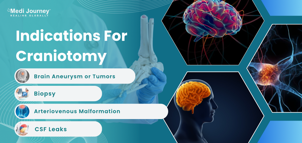
A neurosurgeon will order a craniotomy to treat several brain tumors, such as meningiomas, gliomas, pituitary tumors, schwannomas, and tumors that have spread to the brain from other body parts. The neurosurgeon may also perform a craniotomy to obtain a tissue sample for tumor diagnosis. Other indications of the procedure are:
Before the surgery, the neurosurgeon will explain the procedure to the patient. The patient will also undergo some tests to determine their overall health. These tests will also help determine the craniotomy approach and the dose of anesthetic. The required tests include -
The patient must not eat anything on the day of the surgery. Any additional medicines, such as antibiotics or anticonvulsants, must be taken the day before the surgery.
If the patient is on any blood thinning medication, such as aspirin, the doctor will ask to stop it a few days before surgery.
Like any other surgery, a craniotomy comes with several risks and complications. Some rare complications of a craniotomy include speech difficulties, loss of coordination and balance, difficulty walking, behavioral changes, memory problems, paralysis, and coma.
Some other risks involved with craniotomy are:
Side effects of the procedure depend on several factors, such as the patient's age and overall health, severity of the disease, type of craniotomy, and location of the lesion on the brain.
In a craniotomy procedure, the surgeon removes the skull bone to expose the brain. However, the bone is immediately placed back in place once the surgery is complete. For a craniectomy, the procedure to remove the bone is similar to a craniotomy. The difference is that the bone is not immediately put back after surgery. It is saved to be placed back later, or the surgeon may use an artificial bone. The surgery where the skull bone is replaced is known as cranioplasty.
A craniotomy is a standard procedure to treat brain conditions such as tumors and aneurysms. There are several types and approaches to craniotomy. Patients experiencing symptoms of brain disorders can consult a neurosurgeon to determine the best strategy for them. While a craniotomy is a complex procedure and comes with several risks, new-age treatment options are being continuously developed to minimize the risk factors and improve the outcome of the surgery.
Fill up the form and get assured assitance within 24 hrs!
BDS, Fellowship, MSc
Dr. Ishita Shirvalkar is a dentist, forensic odontologist, and medical writer. She has over two years of clinical experience. She completed her education at reputed institutions such as VSPM Dental College and Research Center in Nagpur.
Head of Department (HOD)
Neurosurgeon
Dr. Anil is a highly experienced Neuro and Spine Surgeon. He has 29+ years of experience and has successfully performed over 10,000 neurosurgical procedures. His expertise lies in Percutaneous Discectomy, Nucleoplasty training, and Minimal Access Spine Su...
Senior Consultant
Medical Oncologist
Nanavati Super Specialty Hospital, Mumbai
WhatsApp UsSenior Director
Gynecologist and Obstetrician, IVF Specialist
Max Super Speciality Hospital, Shalimar Bagh, New Delhi
WhatsApp UsSenior Director
Gynecologist and Obstetrician, IVF Specialist
Max Smart Super Speciality Hospital, Saket, New Delhi
WhatsApp UsSenior Director
Gynecologist and Obstetrician
Max Smart Super Speciality Hospital, Saket, New Delhi
WhatsApp UsSenior Director
Gynecologist and Obstetrician
Max Smart Super Speciality Hospital, Saket, New Delhi
WhatsApp UsSenior Director
Gynecologist and Obstetrician
Max Smart Super Speciality Hospital, Saket, New Delhi
WhatsApp UsThe Art of Effective Communication
 17 December,2025
Read More
 16 December,2025
Read More
 10 December,2025
Read More
 09 December,2025
Read More
 05 December,2025
Read More
 04 December,2025
Read More
Trusted by Patients
"I am Asim from Bangladesh and was looking for treatment in India for neuro. I visited many websites to get the complete information regarding the treatment but I was not satisfied as I was getting confused. In the meanwhile, one of my friends suggested I seek help from Medi Journey as he experienced his medical journey very smoothly and was satisfied with it. They have filtered the top 10 doctors as per experience, the success rate of surgery & profile, so it helps us to choose the best treatment in India. "
"For my knee surgery, Medi Journey guided me to BLK Hospital where I received exceptional care. The team's support and the expertise at BLK Hospital exceeded my expectations. Thank you Medi Journey for making my medical journey stress-free. "
"I came from Iraq for my granddaughter's eye surgery in India facilitated by Medi Journey, due to critical cases they advised us to get a second opinion from the different hospitals before going to surgery. Finally, we went to Fortis Escort Hospital, which helped us to get more confidence for diagnosis. Fortis Escort Hospital has the best eye surgeon team with the latest instruments. Thanks to all team members for providing a high-quality treatment in India at an affordable cost. "
"I came for my hair transplant in India, before coming I was so confused about choosing the best clinic and surgeon for me. But thanks to God one of my friends had a hair transplant in India through Medi Journey. He recommended me to go with them. I am completely happy with my experience with them. They were always very fast in their responses to me. the success rate of my hair transplant surgery is 100%."
"Artemis Hospital, suggested by Medi Journey, turned out to be a great choice for my treatment. The personalized assistance and medical care were exceptional. I'm grateful to Medi Journey for guiding me to a hospital that perfectly matched my needs. Highly recommended! "
"I came from Afghanistan for my treatment in India at Jaypee Hospital, Noida. I had a fantastic experience with Medi Journey. Kudos to them for their incredible support during my medical journey. They not only took care of all the logistics but also connected me with a fantastic healthcare team. Efficient, caring, and highly recommended for a hassle-free medical tourism experience."
"I am Adam from Kano, Nigeria, one of my friends from Nigeria was facilitated by Medi Journey, and he recommended us to go with them. I sent my all reports to them and within 48 hours they reverted with 4 options from different hospitals. They helped me to get a Visa letter from the hospital, arrange pick-up from the airport, and book a hotel for me. Their team is very honest and throughout our stay in India they are with us they are caring for us like his family members. BLK Hospital is the best hospital in India with a top surgical oncologist surgeon team, a very advanced OT, and a Radiotherapy department. I wish more success to Medi Journey. "
"Great experience at the Max Hospital for my spine surgery and was successfully done. I thank my neurosurgeon and his entire team. I recommended all of my country's people to Medi Journey for treatment in India, they choose the best hospital, the best doctors, and the best cost for patients."
"I came to India from Dhaka, Bangladesh for my father-in-law's cardiac surgery at Fortis Hospital. I was confused about choosing the best surgeon for him before coming, but their team helped me to choose the best hospital and best cardiac surgeon in India with very good cost and 100% success rate of surgery. I am very happy with the services, really they make my journey so comfortable that make me feel at home. Thanks again and I like people to choose "Medi Journey" as your travel guide. "
"I am Mohammad from Bangladesh came to India for my general health checkup. Medi Journey offers me the complete package including Pick-up from the airport, hotel services, and 24-hour assistance. They guide you to choose the best hospital in India, the best cost of treatment with top-most doctors and give you complete information about hotel booking, and pick-up from the airport before coming to India They have the best team to help. Always choose Medi Journey for your treatment in India."


