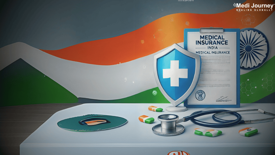
By Country

By City

Enquire now in case of any assistance needed

Peripheral Hemangioma Embolisation Treatment Cost in India

  • Peripheral Hemangioma Embolisation Treatment Cost in India is between

  • Hospital Days:

  • Procedure Duration:

Get Your Estimate Cost

200+
800+
400+
45+

Peripheral hemangioma embolization is a minimally invasive procedure aimed at treating vascular malformations. By using imaging guidance, a catheter is inserted into the blood vessels supplying the hemangioma. Tiny particles or coils are then delivered through the catheter to block the blood flow to the abnormal growth, effectively starving it of oxygen and nutrients. This technique helps reduce the size of the hemangioma, alleviating symptoms such as pain and swelling. Peripheral hemangioma embolization is favored for its low complication rate and quick recovery time, offering a promising solution for patients with symptomatic hemangiomas seeking non-surgical intervention.

Why Do You Need Peripheral Hemangioma Embolization?

Peripheral hemangioma embolization is a crucial procedure employed to manage and alleviate the symptoms associated with peripheral hemangiomas, which are abnormal growths of blood vessels in the peripheral regions of the body. These vascular anomalies often occur in soft tissues such as the skin, muscles, or organs, posing various challenges and risks to the affected individuals. Here's why peripheral hemangioma embolization is necessary:

  • Symptom Relief: Peripheral hemangiomas can cause discomfort, pain, and functional impairment depending on their size and location. Embolization helps alleviate these symptoms by reducing the blood flow to the hemangioma, thereby shrinking its size and relieving associated pressure on nearby structures.
  • Prevention of Complications: Hemangiomas can lead to complications such as bleeding, ulceration, or compression of vital structures. Embolization helps mitigate these risks by reducing the blood supply to the abnormal vessels, thus minimizing the chances of bleeding or other complications.
  • Improved Aesthetic Appearance: Peripheral hemangiomas, especially when located on visible areas of the body, can have a significant impact on one's appearance and self-esteem. Embolization can help improve the cosmetic appearance by reducing the size of the hemangioma and minimizing associated skin changes or deformities.
  • Preservation of Function: In cases where hemangiomas affect organs or tissues critical for function, such as the airway or major nerves, embolization can help preserve function by controlling the growth of the hemangioma and preventing further damage to surrounding structures.
  • Minimally Invasive Approach: Peripheral hemangioma embolization is often performed using minimally invasive techniques, which typically result in faster recovery times, reduced risk of complications, and shorter hospital stays compared to traditional surgical interventions.

Peripheral hemangioma embolization is essential for symptom relief, prevention of complications, improvement of aesthetic appearance, and preservation of function, and is facilitated through minimally invasive procedures, thus offering significant benefits for individuals with peripheral hemangiomas.

Types of Peripheral Hemangioma Embolization

Peripheral hemangioma embolization encompasses various techniques aimed at reducing blood flow to abnormal blood vessels in peripheral regions of the body. These techniques can be broadly classified into several types:

  • Transarterial Embolization (TAE): This is the most common type of embolization for peripheral hemangiomas. In TAE, a catheter is inserted into an artery supplying the hemangioma, and embolic agents such as coils, particles, or liquid embolic are injected to block blood flow to the abnormal vessels.
  • Direct Percutaneous Embolization: In this approach, embolic agents are injected directly into the hemangioma through a needle inserted percutaneously under imaging guidance, bypassing the need for arterial access. This technique is particularly useful for smaller or superficial hemangiomas.
  • Ethanol Embolization: Ethanol can be injected into the blood vessels feeding the hemangioma to induce thrombosis and vascular occlusion. This technique is effective for smaller lesions or in cases where other embolic agents may not be suitable.
  • Laser Photocoagulation: Laser energy is used to heat and coagulate the abnormal blood vessels within the hemangioma, leading to vessel occlusion and shrinkage of the lesion. This technique is often used for superficial hemangiomas, particularly those located in the skin.
  • Radiofrequency Ablation (RFA): RFA involves the use of high-frequency electrical currents to heat and destroy the abnormal blood vessels, causing coagulation and shrinkage of the hemangioma. RFA is typically used for smaller lesions and those located close to the skin surface.

Each type of peripheral hemangioma embolization offers distinct advantages and may be selected based on factors such as the size, location, and characteristics of the hemangioma, as well as patient-specific considerations and the expertise of the interventional radiologist.

How Patients Are Selected For The Procedure? 

The selection of patients for peripheral hemangioma embolization involves a comprehensive evaluation by a multidisciplinary team comprising interventional radiologists, dermatologists, pediatricians (in cases involving pediatric patients), and other specialists as needed. Several factors influence the decision-making process:

  • Clinical Presentation: Patients presenting with symptoms such as pain, functional impairment, bleeding, or cosmetic concerns related to peripheral hemangiomas are prime candidates for embolization.
  • Imaging Studies: Radiological imaging, such as ultrasound, MRI, or CT scans, is essential for assessing the size, location, and vascularity of the hemangioma. Imaging helps determine the feasibility of embolization and guides the selection of appropriate techniques.
  • Vascular Anatomy: Evaluation of the vascular anatomy surrounding the hemangioma is crucial to assess the feasibility and safety of embolization. This includes identifying the arterial supply to the lesion and any nearby vital structures that must be preserved.
  • Patient Characteristics: Age, overall health status, comorbidities, and individual patient preferences are considered when selecting candidates for embolization. For example, pediatric patients with rapidly growing or symptomatic hemangiomas may benefit from early intervention to prevent complications and improve outcomes.
  • Risk-Benefit Assessment: The potential risks and benefits of embolization are carefully weighed for each patient. Factors such as the potential for symptom relief, preservation of function, cosmetic improvement, and the risks of complications associated with the procedure are considered.

The decision to undergo peripheral hemangioma embolization is made collaboratively between the patient, their caregivers (in the case of pediatric patients), and the healthcare team, to achieve the best possible outcome while minimizing risks and addressing the patient's specific needs and concerns.

Risks and Benefits Associated with the Chosen Peripheral Hemangioma Embolization.

Peripheral hemangioma embolization is a procedure that offers several potential benefits for patients with symptomatic or problematic hemangiomas, but it also carries certain risks. Understanding both the risks and benefits is crucial for making informed decisions about undergoing this intervention.

Benefits of Peripheral Hemangioma Embolization

  • Symptom Relief: Embolization can effectively reduce the size of hemangiomas, alleviating symptoms such as pain, discomfort, and functional impairment associated with these vascular anomalies.
  • Cosmetic Improvement: Shrinking the size of the hemangioma through embolization can lead to significant cosmetic improvement, particularly for lesions located in visible areas of the body.
  • Prevention of Complications: By reducing blood flow to the abnormal vessels, embolization can help prevent complications such as bleeding, ulceration, and compression of nearby structures.
  • Preservation of Function: Embolization can preserve the function of vital structures and organs affected by hemangiomas, such as the airway or major nerves, thus preventing functional impairment.
  • Minimally Invasive: Peripheral hemangioma embolization is typically performed using minimally invasive techniques, resulting in shorter recovery times, reduced risk of complications, and shorter hospital stays compared to traditional surgical interventions.

Risks of Peripheral Hemangioma Embolization

  • Bleeding: There is a risk of bleeding at the site of embolization, particularly if the hemangioma is highly vascularized or if adjacent vessels are inadvertently embolized.
  • Infection: Like any invasive procedure, there is a risk of infection at the site of catheter insertion or within the embolized vessels.
  • Tissue Damage: Embolization agents can inadvertently embolize normal tissues or organs, leading to tissue damage or ischemia.
  • Recurrence: While embolization can effectively shrink hemangiomas, there is a risk of recurrence over time, necessitating further interventions.
  • Allergic Reactions: Some embolization agents may cause allergic reactions in rare cases.

Before undergoing peripheral hemangioma embolization, patients and caregivers should discuss these potential risks and benefits with their healthcare providers to make informed decisions tailored to their specific circumstances and treatment goals.

What To Expect After A Peripheral Hemangioma Embolization?

After undergoing peripheral hemangioma embolization, patients can expect a period of recovery and follow-up care to monitor the effectiveness of the procedure and manage any potential complications. Here's what to expect during the post-embolization period:

  • Immediate Recovery: Following the procedure, patients are typically monitored in a recovery area for a brief period to ensure stability before being discharged home or to a hospital room.
  • Pain Management: Patients may experience mild to moderate discomfort or pain at the site of embolization or in the surrounding area. Pain medication may be prescribed to manage discomfort during the initial recovery period.
  • Activity Restrictions: Patients are often advised to limit physical activity and avoid strenuous activities for a certain period following embolization to allow for proper healing. The duration of activity restrictions may vary depending on the specific circumstances of the procedure and the patient's condition.
  • Monitoring: Patients will be scheduled for follow-up appointments with their healthcare provider to monitor the progress of the embolization, assess any changes in symptoms, and address any concerns or complications that may arise.
  • Potential Side Effects: Patients may experience side effects such as bruising, swelling, or numbness at the site of embolization. These side effects are typically temporary and should resolve over time.
  • Long-Term Follow-Up: Long-term follow-up care may be necessary to monitor for recurrence of the hemangioma or any new symptoms or complications. Additional treatments or interventions may be recommended based on the patient's response to embolization and the progression of the condition.
  • Cosmetic Improvement: Over time, patients may notice cosmetic improvement as the size of the hemangioma decreases and associated symptoms resolve. However, the extent of cosmetic improvement can vary depending on factors such as the size and location of the hemangioma and individual healing processes.

The post-embolization period involves close monitoring, symptom management, and follow-up care to ensure optimal outcomes and address any potential complications that may arise. Patients should communicate regularly with their healthcare providers and adhere to recommended follow-up appointments and treatment plans for the best possible recovery.

How Is Peripheral Hemangioma Embolization Performed?

Peripheral hemangioma embolization is a minimally invasive procedure performed by interventional radiologists with specialized training in vascular interventions. Here's an overview of how the procedure is typically performed:

  • Pre-procedure Evaluation: Before the embolization, the patient undergoes a comprehensive evaluation, which may include imaging studies such as ultrasound, MRI, or CT scans to assess the size, location, and vascularity of the hemangioma. The patient's medical history, current medications, and allergies are also reviewed.
  • Anesthesia: Depending on the specific circumstances of the procedure and the patient's preference, local anesthesia or conscious sedation may be administered to ensure comfort during the embolization.
  • Arterial Access: A small incision is made in the skin, typically in the groin area, and a catheter (a thin, flexible tube) is inserted into a large artery, such as the femoral artery, under fluoroscopic guidance. The catheter is then advanced through the arterial system to reach the blood vessels supplying the hemangioma.
  • Angiography: Contrast dye is injected through the catheter to visualize the blood vessels and identify the arterial supply to the hemangioma. This helps the interventional radiologist accurately navigate the catheter to the target vessels.
  • Embolization: Once the target vessels are identified, embolic agents are injected through the catheter to block blood flow to the abnormal vessels feeding the hemangioma. Various embolic materials may be used, including coils, particles, liquid embolic, or sclerosing agents. These agents cause thrombosis (clotting) within the vessels, leading to occlusion and reduction in blood flow to the hemangioma.
  • Post-procedure Care: After embolization, the catheter is removed, and pressure is applied to the insertion site to prevent bleeding. Patients are typically monitored for a brief period in a recovery area before being discharged home or to a hospital room. They may be instructed to limit physical activity and avoid strenuous activities for a certain period following the procedure.
  • Follow-up: Patients are scheduled for follow-up appointments to monitor the progress of the embolization, assess any changes in symptoms, and address any concerns or complications that may arise.

Peripheral hemangioma embolization is a safe and effective procedure for reducing symptoms and improving outcomes in patients with symptomatic or problematic hemangiomas.

Doctors for Peripheral Hemangioma Embolisation in India

Author

Doctor of Pharmacy
Dr. Deepanshu Siwach is a skilled clinical pharmacist with a Doctor of Pharmacy degree. He has 4+ years of experience and has worked with thousands of patients. He has been associated with some of the top hospitals, such as Artemis Gurgaon and Teerthanker

View Profile

Dr. Deepanshu Siwach is a skilled clinical pharmacist with a Doctor of Pharmacy degree. He has 4+ years of experience and has worked with thousands of patients. He has been associated with some of the top hospitals, such as Artemis Gurgaon and Teerthanker...

Frequently Asked Questions

The duration of the procedure varies depending on factors such as the size, location, and complexity of the hemangioma. On average, it may take anywhere from 1 to 3 hours. However, patients should plan for additional time for pre-procedure preparation and post-procedure monitoring in the recovery area.

The success rate of peripheral hemangioma embolization varies depending on factors such as the size, location, and characteristics of the hemangioma, as well as individual patient factors. Generally, embolization is successful in reducing symptoms and shrinking the size of the hemangioma in a significant percentage of cases, with success rates ranging from 70% to 90%.

The recovery time varies depending on factors such as the size and location of the embolized hemangioma and individual healing factors. Generally, patients can resume normal activities within a few days to a week after the procedure, but strenuous activities may be restricted for a longer period.

The effects of peripheral hemangioma embolization can vary. While the procedure itself usually takes 1 to 3 hours, the benefits, such as symptom relief and reduction in hemangioma size, can last for years or even be permanent in some cases. However, individual responses to treatment may vary.

Yes, alternative treatments for peripheral hemangiomas include oral medications, laser therapy, and surgical excision. Oral medications such as beta-blockers or steroids may be prescribed to manage symptoms and shrink the hemangioma. Laser therapy can be used for superficial lesions. Surgical excision may be considered for certain cases, particularly if the hemangioma is large or causing significant functional impairment.

Blogs

The Art of Effective Communication