


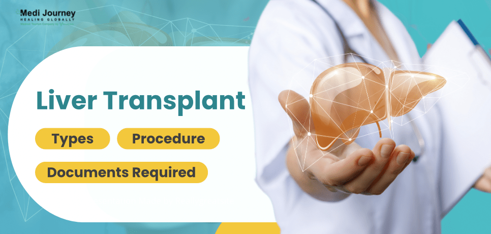
Liver Transplant: Types, Procedure, and Documents Required
 30 September,2024
Read More
30 September,2024
Read More
Enquire now in case of any assistance needed

Contrast Nephrostomy Treatment Cost in India is between

Hospital Days:

Procedure Duration:
Nephrostomy contrast refers to using contrast agents during imaging studies, such as fluoroscopy or CT scans, to visualize the renal collecting system and surrounding structures after percutaneous nephrostomy placement. These contrast agents are injected through the nephrostomy tube into the renal pelvis, allowing healthcare providers to assess the patency of the drainage tract, identify any residual obstruction or leakage, and guide further interventions or treatment planning. Nephrostomy contrast imaging is crucial in monitoring the effectiveness of nephrostomy drainage and ensuring optimal patient outcomes in cases of obstructive uropathy or other urinary tract disorders.
Nephrostomy contrast imaging is necessary to evaluate the patency and functionality of the nephrostomy drainage tract and assess the renal collecting system and surrounding structures. Here's why nephrostomy contrast is essential:
Nephrostomy contrast imaging employs various agents to visualize the renal collecting system and surrounding structures. Here are the common types of nephrostomy contrast:
Selecting patients for nephrostomy contrast imaging involves a comprehensive evaluation by healthcare providers, including radiologists, urologists, and nephrologists. Several factors influence the decision-making process:
Several diagnostic tests and evaluations assess the need for nephrostomy contrast imaging in patients with suspected urinary obstruction or other urinary tract disorders. Here's an overview of the diagnostic modalities commonly utilized:
Nephrostomy contrast imaging offers both risks and benefits that should be carefully considered by patients and healthcare providers:
After undergoing nephrostomy contrast imaging, patients can anticipate several post-procedure experiences:
Nephrostomy contrast imaging is typically performed in a radiology suite or interventional radiology department and involves the following steps:
Senior Consultant
Hepatologist, Medical Gastroenterologist
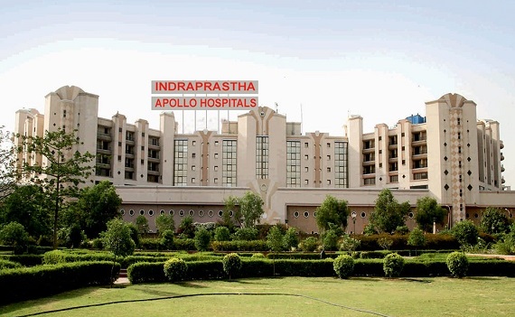
Indraprastha Apollo Hospital, New Delhi
View Doctor Profile Book an AppointmentConsultant
Cardiothoracic and Vascular Surgeon
Apollo Gleneagles Hospital, Kolkata
View Doctor Profile Book an AppointmentSenior Consultant
Hepatologist, Medical Gastroenterologist
Senior Consultant
Gastroenterologist, Hepatologist
Manipal Hospital Formerly AMRI Hospital, Broadway, Kolkata
View Doctor Profile Book an AppointmentDirector
Hepatologist, HPB and Liver Transplant Surgeon, Surgical Gastroenterologist
Aakash Healthcare Super Speciality Hospital, Dwarka, New Delhi
View Doctor Profile Book an AppointmentAssociate Professor
Gastroenterologist, Hepatologist
Doctor of Pharmacy
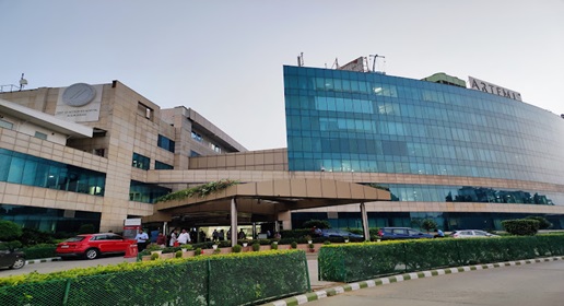
Dr. Deepanshu Siwach is a skilled clinical pharmacist with a Doctor of Pharmacy degree. He has 4+ years of experience and has worked with thousands of patients. He has been associated with some of the top hospitals, such as Artemis Gurgaon and Teerthanker
Dr. Deepanshu Siwach is a skilled clinical pharmacist with a Doctor of Pharmacy degree. He has 4+ years of experience and has worked with thousands of patients. He has been associated with some of the top hospitals, such as Artemis Gurgaon and Teerthanker...
Director
Hepatologist, HPB and Liver Transplant Surgeon, Surgical Gastroenterologist
Aakash Healthcare Super Speciality Hospital, Dwarka, New Delhi
Dr. Ajitabh Srivastava is one of the best Hepatologists, Surgical Gastroenterologists, and Liver Transplant Surgeons in New Delhi. With over 26 years of experience, he has performed over 2500 liver transplant procedures. He specializes in hepato-pancreato-biliary (HPB) surgery, gastrointestinal surgery, acute liver failure treatment, laparoscopic surgery, and gall bladder surgery....
The duration of nephrostomy contrast imaging typically ranges from 30 minutes to an hour. However, the exact time may vary depending on factors such as the complexity of the procedure, the patient's anatomy, and the imaging modalities used. Patients should expect additional time for preparation, recovery, and post-procedure monitoring as needed.
The success rate of nephrostomy contrast imaging is high, with the procedure effectively providing detailed visualization of the renal collecting system and surrounding structures in most cases. Success rates vary depending on factors such as the indication for the procedure, the expertise of the healthcare team, and the patient's anatomical and clinical characteristics. Overall, nephrostomy contrast imaging is considered a valuable diagnostic tool with a high success rate.
After nephrostomy contrast imaging, patients typically have a brief recovery period to monitor for immediate complications. Once stable, patients are usually discharged home. Recovery involves rest and hydration to flush the contrast agent from the body. Patients may experience mild discomfort or pain at the nephrostomy tube insertion site, which can be managed with over-the-counter pain medication as needed.
Patients can typically resume normal activities shortly after nephrostomy contrast imaging, as the procedure is minimally invasive and does not usually require significant recovery time. However, patients may be advised to avoid strenuous activities or heavy lifting for a short period to minimize discomfort or strain on the nephrostomy tube insertion site.
The duration of nephrostomy contrast imaging typically ranges from 30 minutes to an hour. However, the exact length may vary depending on factors such as the complexity of the procedure, the patient's anatomy, and the imaging modalities used. Patients should plan for additional time for preparation, recovery, and any post-procedure monitoring or consultations with healthcare providers.
Yes, alternative imaging modalities and procedures are available to evaluate the renal collecting system and surrounding structures. These alternatives may include ultrasound, CT scans, MRI, or retrograde pyelography. Healthcare providers may recommend alternative imaging modalities or procedures instead of nephrostomy contrast imaging to achieve diagnostic goals depending on the specific clinical scenario and patient factors.
The Art of Effective Communication
30 September,2024
Read More
27 September,2024
Read More
26 September,2024
Read More
25 September,2024
Read More
23 September,2024
Read More
20 September,2024
Read More